
Dental X-rays: types and safety
Dental X-rays show cavities, bone loss, infections, and tooth positions a visual exam cannot see. Modern dental X-rays use very small amounts of radiation. Your dentist chooses the type and timing based on your personal risk.
- 188,364 general dentistry offices listed
- in 51 states
6 min readPublished November 21, 2026Updated November 21, 2026
AI-assisted. AI disclosureEditorial policy
Find General dentist near you












What are dental X-rays?
Dental X-rays are images of your teeth, gums, jawbone, and nearby structures. They show details a regular exam cannot see. For example, they show cavities between teeth, bone loss under the gums, and teeth that have not come in yet.
A small amount of radiation passes through your mouth. It creates a picture on a sensor or film. Hard tissues like teeth and bone absorb more radiation, so they look lighter. Softer tissues and spaces look darker. This contrast helps your dentist see problems early.
Dental X-rays are a common tool for diagnosis and treatment planning. Your dentist uses them with your health history, symptoms, and a clinical exam. Together, these help decide what care may help.
Common types of dental X-rays
Different X-rays show different views. Your dentist chooses the type based on what they need to see.
Bitewing X-rays
Bitewings are one of the most common types. You bite down on a small tab or sensor while the image is taken. They show the upper and lower back teeth in one view. Bitewings help find cavities between teeth and check the bone level around teeth.
Periapical X-rays
A periapical X-ray shows one or two whole teeth from crown to root tip. It also shows the surrounding bone. It helps your dentist see problems near the root, such as infection, an abscess, or bone loss. This type is often used when you have pain in a specific tooth.
Panoramic X-rays
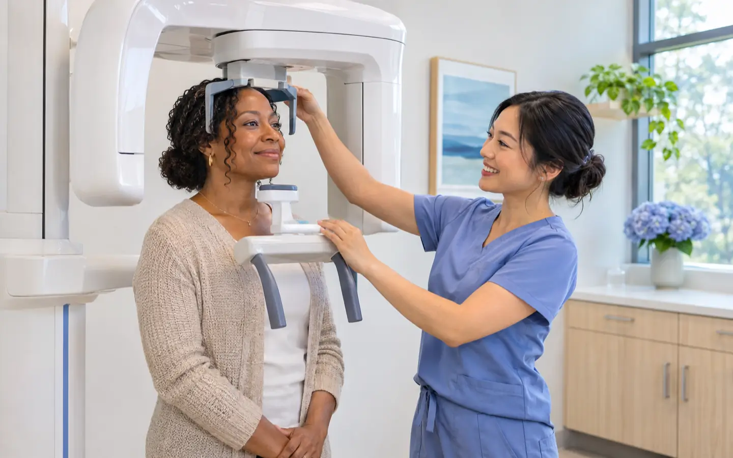
A panoramic X-ray shows the whole mouth in one image. It includes all upper and lower teeth, the jaw joints, sinuses, and wisdom teeth. The machine moves around your head while you stand or sit still. This type helps plan extractions, implants, and orthodontic treatment. It also checks growth and development.
Occlusal X-rays
Occlusal X-rays show the roof or floor of the mouth. They can find extra teeth, teeth that have not broken through the gum, jaw fractures, or cysts. This type is used less often. It can be helpful for children and in certain situations.
Cephalometric X-rays
Cephalometric X-rays show the side of the head. Orthodontists often use them to plan braces or clear aligners. They show the relationship between teeth, jaws, and the facial profile. Not every dental office uses this type.
Cone beam CT
Cone beam CT is a type of 3D imaging. It creates detailed views of teeth, bone, nerves, and soft tissues. Dentists may use it for implant planning, root canal evaluation, or certain surgical procedures. It uses a higher radiation dose than standard dental X-rays. So it is used only when the extra detail is needed.
Why dentists recommend dental X-rays
X-rays can reveal problems before they cause pain or become harder to treat. They may show:
- Cavities between teeth or under existing fillings
- Bone loss from gum disease
- Infection at the tip of a tooth root
- Impacted teeth, such as wisdom teeth
- Cysts, tumors, or other unusual growths
- Tooth and jaw positions for orthodontic planning
- Bone support before implants or dentures
Your dentist may not recommend X-rays at every visit. The decision depends on your age, dental history, current symptoms, and risk for future problems. For example, someone with a history of many cavities may need bitewings more often than someone with stable oral health. Someone with gum disease may need images to check bone levels over time.
How dental X-rays work
During a dental X-ray, a small sensor or piece of film is placed inside your mouth. You may wear a lead apron and a thyroid collar to shield your body. The dental team member steps behind a barrier or out of the room. Then the machine sends a brief, focused beam of radiation. You will not feel anything during the image. The image appears on a computer screen within seconds if digital sensors are used.
Digital X-rays are common in dental offices. They use less radiation than traditional film. They do not require chemical processing. The images can be adjusted on screen. They can also be shared with other providers if needed.
Safety of dental X-rays
Dental X-rays are safe when used appropriately. The radiation in a routine dental X-ray is very small. It is far less than the background radiation you meet in daily life. Still, dental professionals follow the ALARA principle. ALARA means keeping radiation exposure as low as reasonably achievable.
Several steps help keep exposure low:
- Using digital sensors instead of film
- Taking only the images that are needed
- Using lead aprons and thyroid collars when appropriate
- Using rectangular collimation (a way to narrow the X-ray beam)
- Following professional guidelines for timing based on individual risk
Pregnancy and dental X-rays
If you are pregnant or think you may be, tell your dentist. Routine X-rays are often delayed until after birth as a precaution. If you have a dental emergency, such as pain, swelling, or trauma, your dentist may still recommend an X-ray. The benefit of finding the problem may outweigh the very small risk. In that case, shielding can protect your belly and thyroid.
Children and dental X-rays
Children may need X-rays to check for cavities between teeth, monitor growth, or see whether adult teeth are coming in correctly. Dentists use the smallest possible dose. They may take fewer images for children. The timing depends on the child’s cavity risk and development.
How often should you have dental X-rays?
There is no one schedule that fits everyone. Professional dental groups say to decide based on your individual risk, not automatically every year. If your teeth and gums are healthy and your cavity risk is low, you may need X-rays less often.
A crown is a cap that covers a damaged tooth. If you have a history of fillings, crowns, gum disease, dry mouth, or frequent sugary snacks, your dentist may recommend X-rays more often.
Ask your dentist what they are looking for and why an X-ray is recommended. You have the right to understand the reasons.
When dental X-rays may be especially helpful
Certain situations may call for X-rays even if you feel fine:
- Your first visit with a new dentist, to set a baseline
- Before a tooth extraction, root canal, implant, or orthodontic treatment
- After an injury to the mouth, jaw, or face
- When you have tooth pain, swelling, sensitivity, or bleeding gums
- To check healing after a procedure
- To monitor a condition such as gum disease or a cyst
Questions to ask your dentist
You do not need to be nervous about asking questions. Your dental team will explain why an image is needed and what it will show. Consider asking:

Which type of X-ray do you recommend and why?
What are you looking for?
How does my dental history affect how often I need X-rays?
What safety steps do you use to keep radiation low?
Are there alternatives, or can this wait until my next visit?
If I am pregnant or breastfeeding, how does that change the plan?
Your dentist can tailor the plan to your health needs and help you feel comfortable.
Finding a dentist who takes your insurance
If you are looking for a dentist, Dentist Navigator lets you search by insurance, specialty, and location. You can find an office that fits your needs. Call to ask how they handle dental X-rays, what types they use, and how they decide on timing. Many dental offices can give you a cost estimate before your visit. They can also explain what your insurance may cover.
If you are in pain or have a dental emergency, call a dentist right away. For severe swelling, trouble breathing or swallowing, uncontrolled bleeding, or facial trauma, call 911 or go to an emergency room.
 Delta Dental
 Blue Cross Blue Shield
 UnitedHealthcare
 Aetna
 Cigna
 MetLife
 United Concordia
 Guardian
 Medicaid
 Ameritas
 Humana
 Principal
 GEHA
 Careington
 Sun Life
 DentaQuest
 Liberty Dental
 Lincoln Financial
 Medicare Advantage
 TRICARE
 CHIP
 Renaissance
 MCNA
 Spirit
Logos identify plans. No affiliation or endorsement is implied.
Sources
Are dental X-rays safe?
Yes, when used appropriately. Dental X-rays use a very small amount of radiation. Dentists take steps to limit exposure. Talk to your dentist about any concerns.
How often do I need dental X-rays?
There is no one schedule that fits everyone. It depends on your age, risk of decay, gum health, and symptoms. Your dentist may recommend bitewings once a year or less often if your risk is low. Ask about your own situation.
Can I refuse dental X-rays?
You can ask questions and share your concerns. If you decline, your dentist may not see certain problems early. You can work together on a plan. For an emergency or suspected infection, imaging may be important.
What is the difference between a panoramic X-ray and bitewings?
Bitewings show the upper and lower back teeth in one view. They are used to find cavities between teeth. A panoramic X-ray shows the whole mouth, including the jaw joints, sinuses, all teeth, and wisdom teeth in one image.
Are dental X-rays safe during pregnancy?
Routine X-rays are often delayed until after birth as a precaution. If you have a dental emergency, your dentist can use shielding. They can take only needed images. Tell your dentist if you are or may be pregnant.
Related

 Cavities: how they start and how to prevent themCavities are small holes in tooth enamel caused by acid from bacteria that feed on sugars and starches. You can help prevent them with daily brushing and flossing, fluoride, smart eating habits, and regular dental checkups.

 Gum disease: signs and treatment optionsGum disease often starts with bleeding or swollen gums. Early treatment and good daily care can help you manage it and protect your smile.

 How often should you see a dentistMost adults should see a dentist for a checkup and cleaning every 6 months. Your personal schedule may be shorter or longer based on your oral health, medical conditions, and risk factors.
Find a dentist
Search dentists by insurance